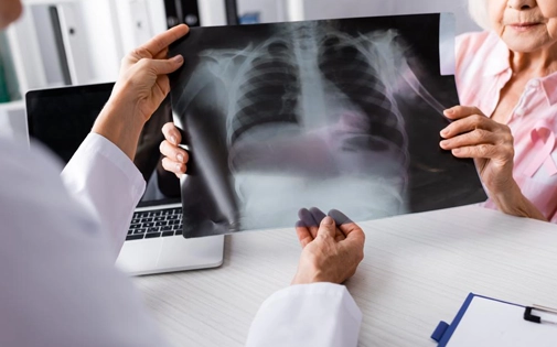
A Chest X-ray (CXR) is a medical imaging test that uses a low dose of ionizing radiation to visualize the structures inside the chest wall. This test allows the examination of the lungs, heart, airways, ribs, and diaphragm. It is a quick, non-invasive diagnostic method used in the evaluation of many chest-related diseases.
A Chest X-ray is commonly used for the diagnosis and follow-up of the following diseases:
No special preparation is required for a Chest X-ray. Jewelry, glasses, or metal items should be removed. The patient may be asked to wear a hospital gown.
A Chest X-ray is usually taken in two different positions:
For patients lying in bed, an Anteroposterior (AP) View is taken. The X-ray machine takes the image from the front of the patient.
The radiology technician will ask the patient to hold their breath and remain still. The X-ray machine briefly emits low-dose radiation to capture the image. The images are processed by a computer and reviewed by a radiologist. The results are sent to your doctor. A Chest X-ray typically takes 5-10 minutes and does not require any pain or recovery time.
The amount of radiation involved in a Chest X-ray is very low and equivalent to daily natural radiation exposure. If you are pregnant, you should inform your doctor. If necessary, protective measures (such as wearing a lead apron) can be taken.
A Chest X-ray is a quick, simple, and effective method for diagnosing diseases related to the lungs, heart, and chest area. It is frequently performed on patients with symptoms like coughing, chest pain, shortness of breath, or fever. The procedure is fast and painless and is considered safe due to its low radiation levels.
A Chest X-ray is one of the fundamental imaging methods used to assess the general condition of the lungs, airways, and chest structures. It can detect many diseases such as infections in the lungs (pneumonia, tuberculosis),tumors, masses, fluid accumulation (pleural effusion),rib fractures, and pulmonary edema. Additionally, chronic diseases (such as COPD, pulmonary fibrosis) or abnormalities in the pleura may also be visualized. However, some small lesions or early-stage diseases may not be detectable with an X-ray and may require further tests.
In a healthy Chest X-ray, the lung tissue should appear clear and symmetrical. Both sides of the lungs should be of equal size with normal air filling. The heart shadow should be of normal size, and the diaphragm domes should be clear and symmetrical. The bronchi, blood vessels, and pleura should be within normal limits. A healthy image would show no opacity (whitening),spots, fluid accumulation, or irregularities. However, as interpreting X-ray films requires expertise, a definitive evaluation should be performed by a radiology specialist.
When having a Chest X-ray, patients are required to remove any clothing covering the chest area or wear a hospital gown. Particularly metal items such as buttons, zippers, bra wires, and jewelry may cause interference in the X-ray image, making diagnosis difficult. Therefore, patients are asked to remove even thin shirts and remain still during the examination. This ensures a clear and accurate image.
A Chest X-ray is requested to evaluate respiratory system symptoms, make a diagnosis, and monitor certain diseases. It is particularly used for investigating infections, tumors, or other lung diseases in patients with symptoms such as prolonged cough, shortness of breath, chest pain, or blood in sputum. It is also frequently used in pre-surgical evaluations, occupational health screenings, and monitoring chronic lung diseases. It plays a crucial role in diagnosing diseases like COVID-19, pneumonia, or tuberculosis.
A Chest X-ray can be performed in state hospitals, private hospitals, radiology centers, and some family health centers. In emergencies, X-rays can be quickly taken in hospital emergency departments, while routine check-ups or doctor-referred procedures can be performed at imaging centers. Additionally, chest X-ray screenings are conducted at some workplaces as part of occupational health and safety measures. After the procedure, the images are typically reviewed by radiology experts, and the relevant doctor may request further tests based on the patient's condition.


Copyright 2026 Prof. Dr. Elif Küpeli. All rights reserved.
Prof. Dr. Elif KüpeliChest Diseases Specialist