 +90 539 716 24 01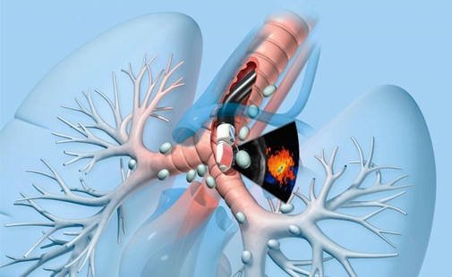
Endobronchial Ultrasound (EBUS) is a diagnostic procedure that uses a bronchoscope equipped with an ultrasound probe to visualize lymph nodes surrounding the airways. It is primarily used for the diagnosis and staging of lung cancer. In addition, it is also helpful in diagnosing other diseases that cause lymph node enlargement, such as tuberculosis, sarcoidosis, and lymphoma.
EBUS combines bronchoscopy and ultrasound, allowing both direct visual examination of the airways and the ability to obtain tissue samples (biopsy) from deep structures. This enables highly accurate diagnosis without the need for surgical procedures.
The EBUS device is a specialized bronchoscope with a high-frequency ultrasound probe at its tip. This system detects enlarged lymph nodes or pulmonary nodules around the airways and provides real-time imaging on a monitor.
The EBUS probe also allows fine-needle aspiration biopsy (EBUS-TBNA) from lymph nodes. In this way, the physician can directly sample suspicious areas and send them for pathological examination.
EBUS is performed under general anesthesia or deep sedation. The patient is placed in a supine position, and the bronchoscope is advanced through the mouth into the airways.
Once the device reaches the bronchial tree, surrounding lymph nodes and tissues are carefully examined using the ultrasound probe. When suspicious areas are identified, a biopsy is obtained using a special needle.
The procedure usually takes 45–60 minutes, and the patient is observed for a short period afterward. Hospitalization is generally not required.
EBUS is mainly used in the following conditions:
Thanks to these features, EBUS plays a critical role in pulmonary medicine and thoracic oncology.
Before EBUS, the patient must be fasting, usually for 6–8 hours. If the patient is using blood-thinning medications, these should be temporarily discontinued according to the physician’s instructions. During the procedure, heart rate, oxygen saturation, and blood pressure are continuously monitored. Due to anesthesia, the patient does not feel pain or discomfort. Possible allergies and bleeding risks must be carefully evaluated beforehand.
Compared to conventional bronchoscopy, CT (computed tomography),or PET scans, EBUS provides more detailed anatomical information. Its greatest advantage is the ability to perform real-time, ultrasound-guided biopsies. It also allows lymph node diagnosis without the need for surgical intervention. This offers the patient lower risk, faster recovery, and reduced cost.
Biopsy samples obtained via EBUS are examined microscopically in the pathology laboratory. This analysis helps determine cell type, presence of malignancy, or signs of infection. The diagnostic accuracy of EBUS exceeds 90%, especially in lung cancer staging. Therefore, it is recommended in many international guidelines as the first-choice method for mediastinal evaluation.
EBUS is generally a safe procedure, and complication rates are very low. However, in rare cases, the following side effects may occur:
These complications are usually temporary and treatable.
EBUS bronchoscopy typically takes about 45 to 60 minutes, depending on the extent of the examination and the number of biopsies taken. After the procedure, the patient is observed for about one hour and can usually be discharged the same day. It is recommended not to drive and to rest until the effects of anesthesia wear off.
EBUS is commonly requested for the diagnosis or staging of lung cancer, evaluation of lymph node enlargement, or diagnosis of infectious and granulomatous diseases. It allows tissue sampling without surgical intervention and helps accurately determine disease spread.
EBUS is performed by the Department of Pulmonology (Chest Diseases). In some centers, it may also be carried out by Thoracic Surgery or Interventional Pulmonology units. The procedure is done in a bronchoscopy suite or operating room using specialized EBUS equipment, usually by an experienced pulmonologist.





Copyright 2026 Prof. Dr. Elif Küpeli. All rights reserved.

 Prof. Dr. Elif KüpeliPulmonary Diseases, Pulmonologist
 Prof. Dr. Elif Küpeli
 Türkçe
 English
 +90 539 716 24 01